

Cone Beam Imaging in Metairie, LA
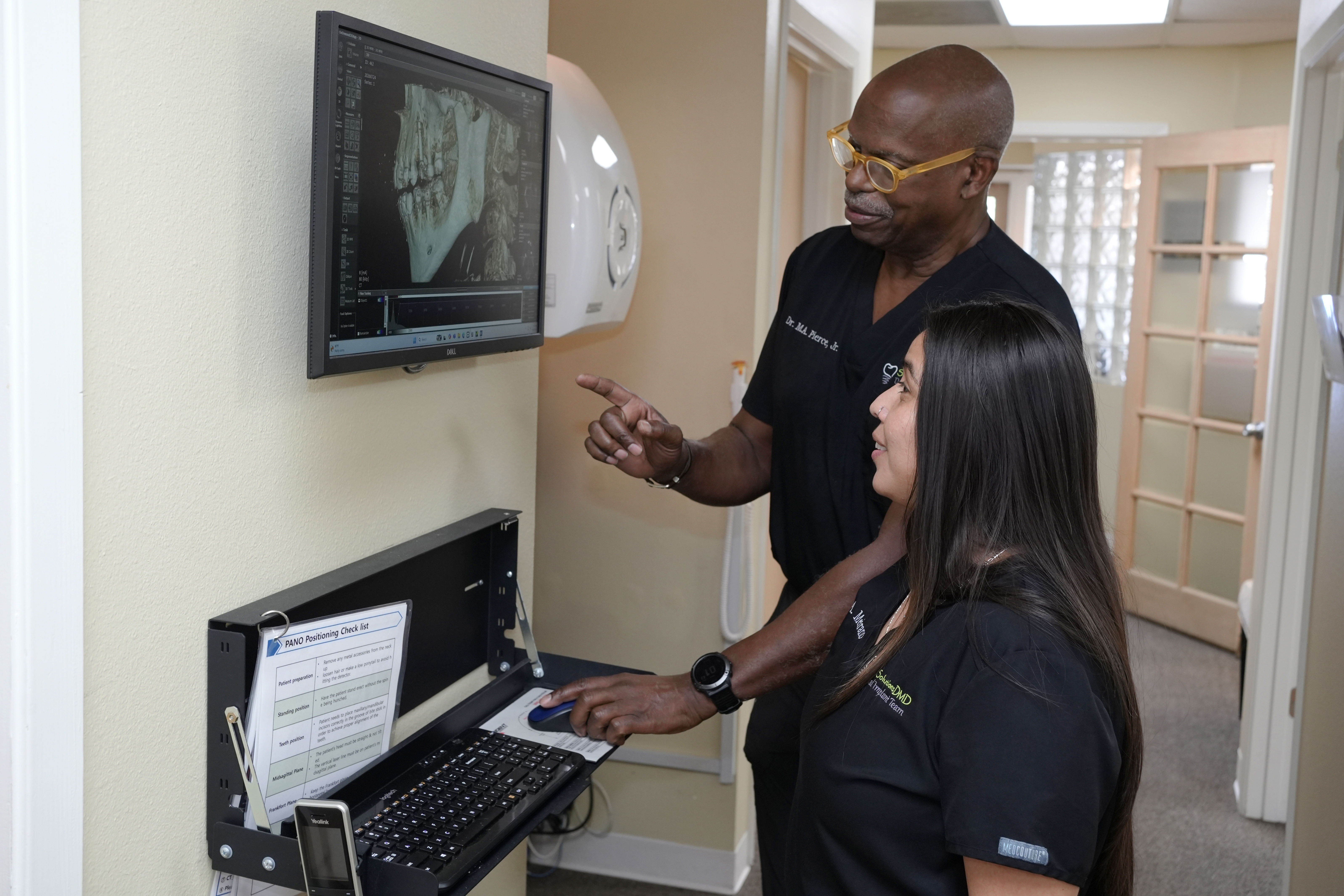
Cone beam imaging in Metairie, Louisiana, provides detailed 3D views of teeth, jaws, and facial structures to support precise diagnosis and treatment planning. Often called cone beam CT (CBCT), this dental technology produces a comprehensive image in a single scan, helping patients and clinicians see what standard two-dimensional dental X-rays may miss.
Cone Beam Imaging Explained
Cone beam imaging uses a cone-shaped beam of X-rays to capture hundreds of images as the scanner rotates around your head. Software reconstructs these images into a 3D model of your teeth, bone, nerves, and sinuses. This allows a clear look at anatomy from any angle. Compared with a medical CT, CBCT is optimized for dentistry and generally uses a lower radiation dose. Compared with panoramic or bitewing X-rays, CBCT shows far more detail, including the exact location of tooth roots and vital structures.
CBCT is often recommended when two-dimensional images are not enough to answer a clinical question. Common reasons include dental implant planning, complex root canal (endodontic) cases, impacted teeth, wisdom teeth evaluation, temporomandibular joint (TMJ) concerns, jaw pathology, orthodontic assessment, and airway evaluation. Patients frequently ask what cone beam imaging is used for, how CBCT works, and the benefits of 3D dental X-rays. This page addresses those questions in a clear, step-by-step way.
A Three-Dimensional View
Cone beam imaging produces a three-dimensional scan of teeth, bone, and surrounding structures. Standard X-rays are flat, so depth and volume are difficult to judge.
For implant planning in particular, seeing bone height and width in three dimensions makes it possible to plan position precisely before any treatment begins.
Why Consider Cone Beam Imaging?
- 3D detail for confident diagnosis and treatment planning.
- Accurate assessment of bone volume and density for dental implants.
- Clear visualization of nerves, roots, and sinuses to reduce complications.
- Targeted evaluation that may prevent surprise findings during procedures.
- Efficient imaging that typically takes less than a minute.
- Lower radiation dose than a medical CT while providing rich detail.
Planning Implant Treatment?
Three-dimensional imaging helps determine whether there is enough bone to support an implant.
Better Planning, Fewer Surprises
Detailed imaging means treatment can be mapped out in advance, which supports safer, more predictable care and a clearer conversation about your options.
- Used when standard X-rays are not enough
- Supports implant and complex treatment planning
- Findings reviewed with you before decisions
The Cone Beam Imaging Process
Before Your Scan
Preparation is simple. You will remove glasses, earrings, hairpins, and removable dental appliances. No special diet or medication changes are needed for most patients. If you are or may be pregnant, inform the team so timing and necessity can be reviewed.
During The Scan
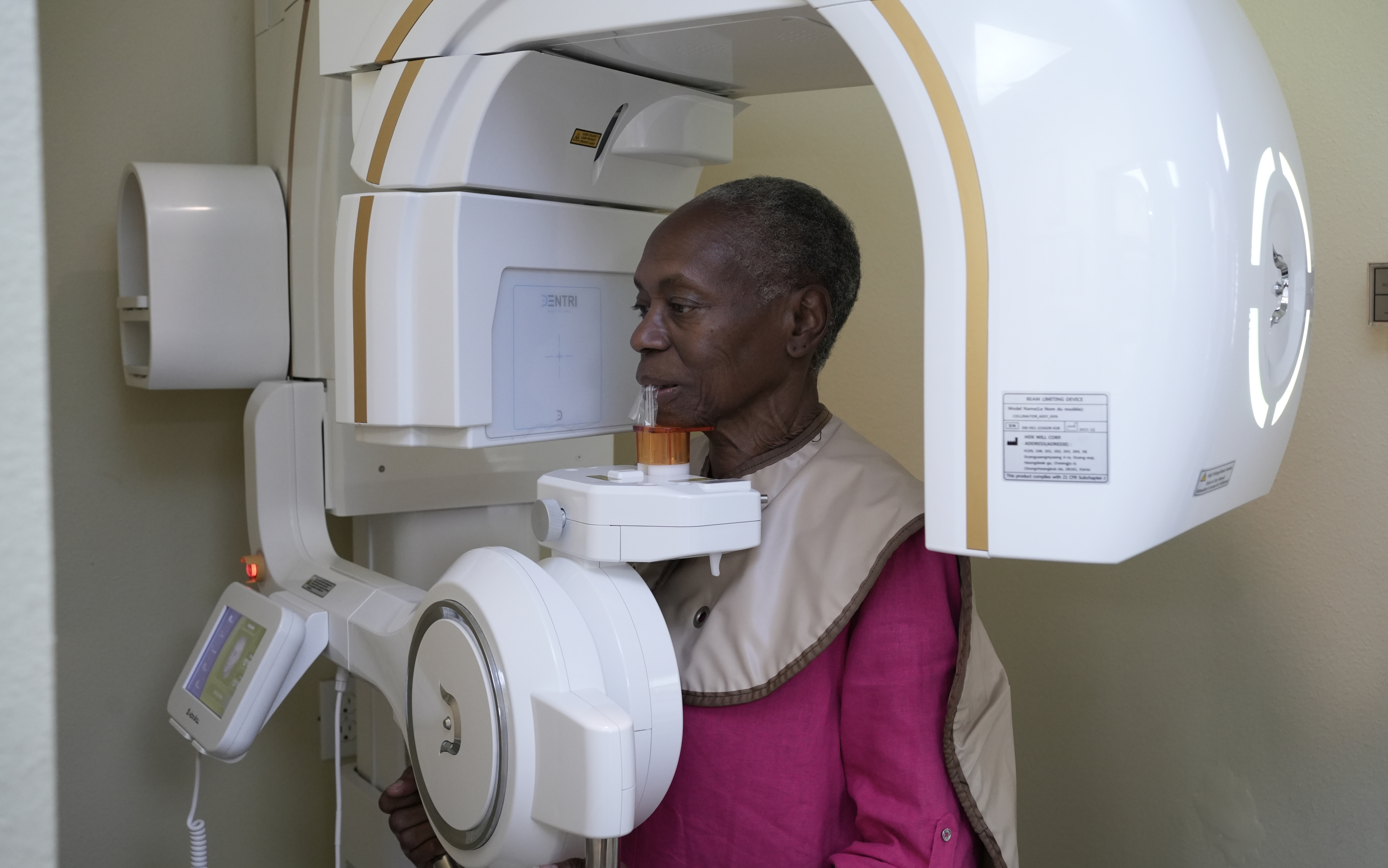
You will stand or sit while the machine gently rotates around your head. The scan itself often takes 10 to 20 seconds. You can breathe normally and there is no contact with the scanner other than a small support to help you stay still. The process is quiet and does not cause pain.
After The Scan
The 3D images are available almost immediately. They can be viewed from many angles, with cross-sections that show precise measurements. This helps explain findings in plain terms and supports choices such as whether bone grafting is needed before an implant.
What to Expect
Most appointments take only a few minutes beyond normal visit time. A lead apron and, when appropriate, a thyroid collar are used for protection. CBCT uses ionizing radiation, so scans are taken only when the expected benefit outweighs the risk. In many cases, a single CBCT can replace multiple traditional X-rays by answering several questions at once.
Metal objects such as braces, implants, or root canal fillings can create minor artifacts on the image. Modern software reduces these, and they rarely limit the dentist's ability to interpret the scan. Results are reviewed with you chairside so you understand the diagnosis and the plan. When helpful, Dr. Mitchell Pierce can coordinate with your specialist using the same 3D data set.
Understanding Your CBCT Options
CBCT fields of view range from small (a few teeth) to large (entire jaws and sinuses). The selected view is tailored to the clinical question to keep radiation as low as reasonably achievable. Many patients appreciate how CBCT clarifies issues like the position of an impacted canine, the shape of a curved root, or the relationship of an implant site to the nerve canal. These insights support safer care and fewer surprises.
Schedule a Consultation
Contact Smile Solutions DMD at 504-613-5100 to schedule a visit and discuss your treatment.

Frequently Asked Questions About Cone Beam Imaging
Helpful Answers to Common Questions
Yes. CBCT uses a lower dose than a medical CT and is used only when the diagnostic value justifies exposure. Shielding and focused fields of view further limit dose.
CBCT is designed for dentistry, offers excellent bone and tooth detail, and typically uses a lower dose than medical CT. It shows far more 3D detail than a panoramic X-ray.
No. The scan is open, fast, and silent. You stand or sit in the unit, and nothing touches your face except a positioning guide.
CBCT is often used for dental implants, complex root canals, impacted or wisdom teeth, TMJ concerns, orthodontic assessment, and evaluating sinuses or airway.
Yes, when there is a clear clinical need. Settings and field of view are adjusted to minimize exposure for growing patients.
Imaging is generally deferred during pregnancy unless urgent. Always inform the dental team so risks and benefits can be considered.