

Digital X-Rays in Metairie, LA
Digital X-rays at Smile Solutions DMD help patients receive precise, timely diagnoses with far less radiation than traditional film. This page explains how digital dental radiographs work, when they are used, and what to expect at an appointment. The goal is to make decisions easier by presenting clear, practical information about digital X-rays in Metairie, Louisiana.
Digital X-Rays Explained
Digital X-rays use electronic sensors instead of film to capture images of your teeth, roots, and supporting bone. The sensor sends the image to a computer in seconds, where the dental team can adjust contrast, magnify areas, and compare findings over time. Many patients ask a common question: What are digital X-rays? In simple terms, they are diagnostic images that guide preventive care, detect decay between teeth, assess bone levels, and evaluate existing restorations.
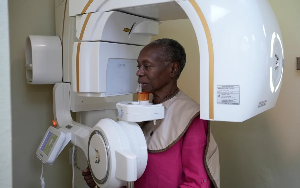
Common views include bitewing images that show the crowns of back teeth and the bone between them, and periapical images that show an entire tooth from crown to root. A panoramic image can display the entire jaw and is helpful for wisdom teeth, growth patterns, and jaw joints. In some cases, a cone beam CT scan provides 3D details for complex treatment planning. These tools support early detection and help answer how digital X-rays work in everyday care while maintaining a strong focus on dental radiographs safety.
Seeing What an Exam Cannot
Digital X-rays capture detailed images of tooth structure and supporting bone, showing decay between teeth and changes that are not visible during a visual exam.
Images appear on screen immediately, so we can review them with you and explain exactly what we are seeing before recommending anything.
Benefits of Digital X-Rays
- Lower radiation exposure compared with traditional film in most cases.
- Instant images that speed up diagnosis and reduce time in the chair.
- High-resolution detail that helps reveal early decay and infection.
- Image enhancement tools that improve accuracy and comparison over time.
- Easy electronic sharing for second opinions or specialist care when needed.
- No chemical processing, which is more environmentally friendly.
Questions About Dental Imaging?
We are happy to explain what we look for and why imaging is recommended in your case.
Imaging Only When It Helps
How often X-rays are taken depends on your cavity risk, gum health, and history rather than a fixed schedule. We take images when they will genuinely inform your care.
- Frequency based on your individual risk
- Images explained in plain language
- Records kept for comparison over time
How Digital X-Rays Work
- Preparation: A small sensor or a panoramic chin rest is positioned comfortably.
- Positioning: The team aligns the sensor and X-ray beam to the specific teeth or jaw area.
- Exposure: A quick, low-dose pulse captures the image in a fraction of a second.
- Review: The image appears on-screen for evaluation, zooming, and side-by-side comparison.
- Discussion: Findings are explained with visuals so you can see areas of concern.
What to Expect
Most digital X-rays take only a few minutes. Sensors may feel bulky at first, but adjustments and cushioning can improve comfort. A lead apron and, when appropriate, a thyroid collar are used to minimize scatter. The dental team recommends an interval based on your history and risk for cavities or gum disease. Low-risk adults may need bitewings every 12–24 months, while others benefit from more frequent checks. If you are pregnant or think you may be, inform the office so imaging can be timed and shielded appropriately.
Patients often ask about dental X-ray radiation dose. Digital systems typically use significantly less radiation than film, and exposures are kept as low as reasonably achievable while still producing diagnostic images.
Schedule Your Appointment
Contact Smile Solutions DMD at 504-613-5100 to schedule an exam.

Frequently Asked Questions About Digital X-Rays
Helpful Answers to Common Questions
Yes. Digital systems use a low radiation dose, often lower than traditional film. A lead apron and careful technique limit scatter. For most patients, the benefit of detecting hidden problems early outweighs the minimal exposure.
Frequency depends on your oral health and risk level. New patients often need a baseline set. After that, intervals commonly range from 6–24 months for bitewings, with periapical or panoramic images taken as needed to assess symptoms or monitor treatment.
Bitewing X-rays help find decay between back teeth and evaluate bone levels. Periapical views show roots and surrounding bone. Panoramic images capture the entire jaw. In select cases, a cone beam CT scan provides 3D information for complex planning.
No. The exposure is painless. Some people feel brief pressure from the sensor. Let the team know if a spot is sensitive; small adjustments, cushioning, or a different sensor size can increase comfort.
Dental emergencies may still require imaging with shielding. Non-urgent X-rays are often postponed until after pregnancy. Always tell the office and coordinate care with your physician so timing and protection are optimized.
Panoramic images show the entire jaw in one view and are useful for wisdom teeth, growth, and jaw joints. Bitewing images are focused snapshots of back teeth that excel at detecting decay between teeth and tracking bone height. For questions or to plan your next checkup with digital X-rays in [city], [st], contact [practice_name] at <a href="tel:[phone]">[phone]</a> and schedule an appointment with Dr. [doctor_name].